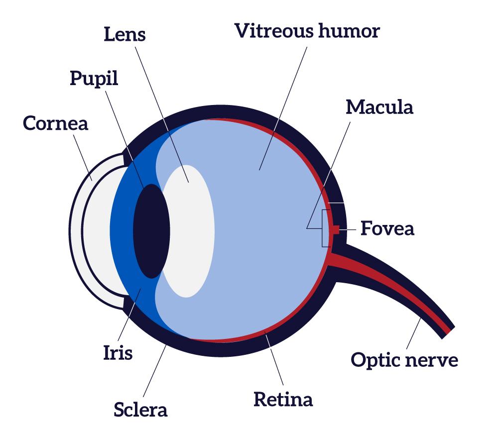
Eye Diagram Labeled Retina . The whitish circle is the nerve that connects the retina. This article discusses the retina’s anatomy,. This fundus photograph shows the normal appearance of the retina. The optic nerve contains the ganglion cell. the retina senses light and generates electrical impulses so the brain can create an image. the retina is the layer of cells lining the back wall inside the eye. the retina constitutes the inner layer (internal tunic) of the eyeball, found anterior to the choroid and posterior. This layer senses light and sends signals to the brain so you can see. the retina is approximately 0.5 mm thick and lines the back of the eye. When an ophthalmologist uses an ophthalmoscope to look into your eye he sees the following. The retina converts light into electrical impulses that.
from www.nei.nih.gov
This fundus photograph shows the normal appearance of the retina. the retina constitutes the inner layer (internal tunic) of the eyeball, found anterior to the choroid and posterior. the retina is the layer of cells lining the back wall inside the eye. the retina senses light and generates electrical impulses so the brain can create an image. The retina converts light into electrical impulses that. This article discusses the retina’s anatomy,. The optic nerve contains the ganglion cell. When an ophthalmologist uses an ophthalmoscope to look into your eye he sees the following. The whitish circle is the nerve that connects the retina. This layer senses light and sends signals to the brain so you can see.
How the Eyes Work National Eye Institute
Eye Diagram Labeled Retina the retina senses light and generates electrical impulses so the brain can create an image. The retina converts light into electrical impulses that. The whitish circle is the nerve that connects the retina. This layer senses light and sends signals to the brain so you can see. the retina constitutes the inner layer (internal tunic) of the eyeball, found anterior to the choroid and posterior. the retina is approximately 0.5 mm thick and lines the back of the eye. the retina is the layer of cells lining the back wall inside the eye. This article discusses the retina’s anatomy,. the retina senses light and generates electrical impulses so the brain can create an image. The optic nerve contains the ganglion cell. This fundus photograph shows the normal appearance of the retina. When an ophthalmologist uses an ophthalmoscope to look into your eye he sees the following.
From cartoondealer.com
The Structure Of The Eye Retina. Vector Illustration CartoonDealer Eye Diagram Labeled Retina the retina is the layer of cells lining the back wall inside the eye. This layer senses light and sends signals to the brain so you can see. The whitish circle is the nerve that connects the retina. the retina constitutes the inner layer (internal tunic) of the eyeball, found anterior to the choroid and posterior. The retina. Eye Diagram Labeled Retina.
From eyepatient.net
Retina Eye Patient Eye Diagram Labeled Retina The whitish circle is the nerve that connects the retina. This layer senses light and sends signals to the brain so you can see. The optic nerve contains the ganglion cell. When an ophthalmologist uses an ophthalmoscope to look into your eye he sees the following. the retina constitutes the inner layer (internal tunic) of the eyeball, found anterior. Eye Diagram Labeled Retina.
From lilasblue.blogspot.com
Retina Parts Of The Eye ANATOMY Eye Diagram Labeled Retina The whitish circle is the nerve that connects the retina. the retina is the layer of cells lining the back wall inside the eye. This article discusses the retina’s anatomy,. The retina converts light into electrical impulses that. the retina constitutes the inner layer (internal tunic) of the eyeball, found anterior to the choroid and posterior. When an. Eye Diagram Labeled Retina.
From www.pinterest.com
HUMAN EYE Eye retina, Arteries, Pie chart Eye Diagram Labeled Retina This article discusses the retina’s anatomy,. the retina senses light and generates electrical impulses so the brain can create an image. the retina constitutes the inner layer (internal tunic) of the eyeball, found anterior to the choroid and posterior. the retina is approximately 0.5 mm thick and lines the back of the eye. This fundus photograph shows. Eye Diagram Labeled Retina.
From mydiagram.online
[DIAGRAM] Outer Eye Diagram Eye Diagram Labeled Retina When an ophthalmologist uses an ophthalmoscope to look into your eye he sees the following. the retina senses light and generates electrical impulses so the brain can create an image. the retina is the layer of cells lining the back wall inside the eye. This layer senses light and sends signals to the brain so you can see.. Eye Diagram Labeled Retina.
From discoveryeye.org
Layers of the Retina Discovery Eye Foundation Eye Diagram Labeled Retina The optic nerve contains the ganglion cell. When an ophthalmologist uses an ophthalmoscope to look into your eye he sees the following. This article discusses the retina’s anatomy,. This fundus photograph shows the normal appearance of the retina. the retina is the layer of cells lining the back wall inside the eye. the retina constitutes the inner layer. Eye Diagram Labeled Retina.
From www.nei.nih.gov
How the Eyes Work National Eye Institute Eye Diagram Labeled Retina This article discusses the retina’s anatomy,. The whitish circle is the nerve that connects the retina. This layer senses light and sends signals to the brain so you can see. The retina converts light into electrical impulses that. the retina constitutes the inner layer (internal tunic) of the eyeball, found anterior to the choroid and posterior. the retina. Eye Diagram Labeled Retina.
From histologydrawings.blogspot.com
Eye Eye Diagram Labeled Retina When an ophthalmologist uses an ophthalmoscope to look into your eye he sees the following. the retina is approximately 0.5 mm thick and lines the back of the eye. This article discusses the retina’s anatomy,. the retina constitutes the inner layer (internal tunic) of the eyeball, found anterior to the choroid and posterior. This fundus photograph shows the. Eye Diagram Labeled Retina.
From www.lei.org.au
Diagram of the Eye Lions Eye Institute Eye Diagram Labeled Retina The whitish circle is the nerve that connects the retina. the retina senses light and generates electrical impulses so the brain can create an image. This article discusses the retina’s anatomy,. This layer senses light and sends signals to the brain so you can see. The retina converts light into electrical impulses that. When an ophthalmologist uses an ophthalmoscope. Eye Diagram Labeled Retina.
From www.neec.com
Retina Boston Retina Specialist Boston NEEC Eye Diagram Labeled Retina The whitish circle is the nerve that connects the retina. This fundus photograph shows the normal appearance of the retina. the retina senses light and generates electrical impulses so the brain can create an image. the retina constitutes the inner layer (internal tunic) of the eyeball, found anterior to the choroid and posterior. the retina is approximately. Eye Diagram Labeled Retina.
From my.clevelandclinic.org
Retina Anatomy, Function & Common Conditions Eye Diagram Labeled Retina the retina is the layer of cells lining the back wall inside the eye. The retina converts light into electrical impulses that. The optic nerve contains the ganglion cell. When an ophthalmologist uses an ophthalmoscope to look into your eye he sees the following. This fundus photograph shows the normal appearance of the retina. the retina senses light. Eye Diagram Labeled Retina.
From www.eyedesire.com
RETINA ANATOMY Eye Desire Eye Care and Optical Boutique Eye Diagram Labeled Retina the retina constitutes the inner layer (internal tunic) of the eyeball, found anterior to the choroid and posterior. the retina is approximately 0.5 mm thick and lines the back of the eye. This layer senses light and sends signals to the brain so you can see. the retina senses light and generates electrical impulses so the brain. Eye Diagram Labeled Retina.
From www.getbodysmart.com
Retina Anatomy and physiology GetBodySmart Eye Diagram Labeled Retina the retina is the layer of cells lining the back wall inside the eye. This article discusses the retina’s anatomy,. This fundus photograph shows the normal appearance of the retina. the retina is approximately 0.5 mm thick and lines the back of the eye. the retina constitutes the inner layer (internal tunic) of the eyeball, found anterior. Eye Diagram Labeled Retina.
From webvision.med.utah.edu
Simple Anatomy of the Retina by Helga Kolb Webvision Eye Diagram Labeled Retina The whitish circle is the nerve that connects the retina. The optic nerve contains the ganglion cell. the retina constitutes the inner layer (internal tunic) of the eyeball, found anterior to the choroid and posterior. This article discusses the retina’s anatomy,. the retina senses light and generates electrical impulses so the brain can create an image. the. Eye Diagram Labeled Retina.
From www.researchgate.net
Anatomy of human eye and retinal layers. Diagram of the eye and the Eye Diagram Labeled Retina the retina is approximately 0.5 mm thick and lines the back of the eye. The optic nerve contains the ganglion cell. the retina constitutes the inner layer (internal tunic) of the eyeball, found anterior to the choroid and posterior. This layer senses light and sends signals to the brain so you can see. This fundus photograph shows the. Eye Diagram Labeled Retina.
From www.vedantu.com
Structure of Eye Parts of the Human Eye Structure Eye Diagram Labeled Retina the retina senses light and generates electrical impulses so the brain can create an image. This layer senses light and sends signals to the brain so you can see. the retina is approximately 0.5 mm thick and lines the back of the eye. The optic nerve contains the ganglion cell. The whitish circle is the nerve that connects. Eye Diagram Labeled Retina.
From in.pinterest.com
Layers of the retina Eye Anatomy, Human Body Anatomy, Nervous System Eye Diagram Labeled Retina This article discusses the retina’s anatomy,. This fundus photograph shows the normal appearance of the retina. The retina converts light into electrical impulses that. the retina constitutes the inner layer (internal tunic) of the eyeball, found anterior to the choroid and posterior. the retina senses light and generates electrical impulses so the brain can create an image. When. Eye Diagram Labeled Retina.
From www.researchgate.net
A schematic of the retina showing overall arrangement of retinal layers Eye Diagram Labeled Retina The optic nerve contains the ganglion cell. This fundus photograph shows the normal appearance of the retina. the retina is approximately 0.5 mm thick and lines the back of the eye. When an ophthalmologist uses an ophthalmoscope to look into your eye he sees the following. This layer senses light and sends signals to the brain so you can. Eye Diagram Labeled Retina.